


Figure 2.
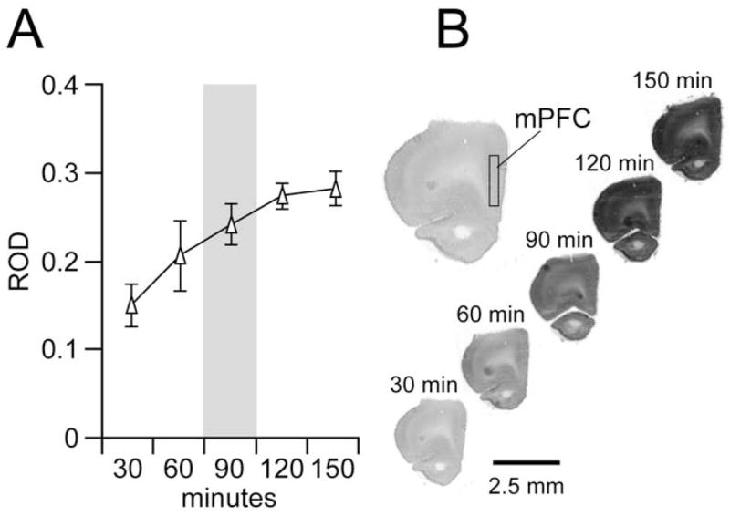
(A) Relationship between incubation time and prefrontal cortex (PFC) cytochrome oxidase I (CO-I) levels in slides containing normal nonstimulated brain sections (mean ± SD; n = 4; 5– 6 sections per animal). After incubating for 120 –150 minutes, PFC CO-I staining reached saturation level. As indicated in gray, 90 minutes of incubation was the most appropriate time point at which to assess the effect of neonatal ventral hippocampal lesion and ventral tegmental area stimulation on PFC CO-I activity. The variability of CO-I staining was lower than that from 60 minutes, and therefore it allows a better relative optical density range to detected CO-I changes in the PFC and other cortical and subcortical regions. (B) Representative examples of coronal forebrain sections at the level of the PFC showing CO-I staining after 30, 60, 90, 120, and 150 minutes of incubation.