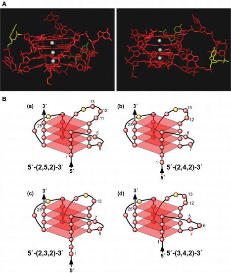
Figure 9.
(A) Model of the biologically relevant NHEPDGF-A G-quadruplex [loop isomer 5′-(2,5,2)-3′], which contains two 2-base double-chain reversal loops and one 5-base intervening loop (guanines = red, cytosines = yellow and K+ ions = white). For clarity, hydrogen atoms have been removed. In the left panel, the two 2-base double-chain reversal loops are shown on each side of model, and in the right panel, the model has been rotated to show the 5-base intervening loop on the right side of model. (B) Proposed folding patterns of the four different loop isomers formed in the core sequence of NHEPDGF-A. Guanines = red, cytosines = yellow.

