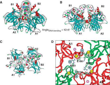
Figure 3.
Structural details of the potential H14 binding mode, in which two swapped copies of p53 dimer bind DNA symmetrically in respect to full-site palindrome. p53-tetramer–Puma BS2 complex is used here. Chains binding DNA specifically at quarter-sites one and four are the B chains in the p53-trimer–DNA complex. B1 and B2 are used to term their position in two dimers, respectively. The A chains assist in the DNA recognition and provide tetramer stability. A1 and A2 are used to name the chain position. (A) A view along the DNA chain. (B) A view perpendicular to the DNA chain, in which the p53 tetramer is inside the DNA loop. (C) A view from the top of the complex illustrating the arrangement of the core domain tetramer. (D) Atomic details of the salt bridges stabilizing the p53 tetramer.

