
Figure 3.
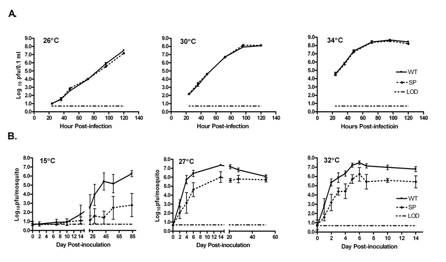
Comparison of growth of wild type (WT) and a small plaque (SP) variant WNV in vitro in a mosquito cell line and in vivo in mosquitoes. (A) C6/36 cells in 6-well plates were infected with Vero cell amplified WT or SP stock virus in triplicate at an MOI of 0.01 pfu/cell. Virus was harvested from the medium at the indicated time points and titers determined by plaque assay on Vero cells. Symbols represent the mean titer and standard deviation for each time point. The limit of detection (LOD) was 0.7 log10pfu/0.1ml of supernatant of infected cell cultures; in cases where the sample had no detectable virus, a titer of 0.65 log10pfu/0.1ml was used for calculations. (B) Cx. pipiens mosquitoes were inoculated intrathoracically at 10× the ID50 of WT (5 pfu per mosquito) or SP (21 pfu per mosquito) with Vero cell amplified WT or SP stock virus, and were maintained at the indicated temperatures. Five mosquitoes per virus were harvested immediately following inoculation, and 10 mosquitoes were harvested at each time point thereafter. Viral titers in the mosquito bodies were determine by plaque titration on Vero cells. Symbols represent the mean viral titer and standard deviation for each time point. The limit of detection (LOD) was 0.7 log10pfu/mosquito, and in cases where the sample had no detectable virus, a titer of 0.65 log10pfu/mosquito was used for calculations.