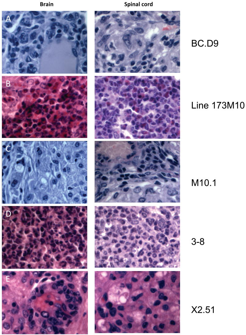
Figure 1.
Inflammatory infiltrates in CNS tissues of T cell recipients. Representative hematoxylin and eosin stained sections from brain and spinal cord of indicated T cell clone or line recipients are shown, magnified x600. All mice received 10 × 106 in vitro activated T cells intravenously. Mice were sacrificed within 1–2 days of showing clinical signs of disease. (A) Brain and spinal cord of BCD9 recipient sacrificed 10 days after transfer. Infiltrates consist primarily of macrophages and lymphoid cells, and are concentrated in perivascular and meningeal regions. (B) Brain and spinal cord of line 173M10 recipient 10 days after transfer. Abcesses of neutrophils and eosinophils are present in both brain and spinal cord tissues. (C) Tissues from clone M10.1 recipient 8 days after transfer; macrophages dominate among infiltrating cells. (D) Lateral medulla is completely taken over by infiltrating neutrophils in 3–8 recipients, 10 days after transfer. Spinal cord lesions are rare and isolated, but also consist of very large foci of neutrophils. (E) Tissues from X2.51 recipient 10 days post transfer, dominated by foamy macrophages and lymphoid cells.

