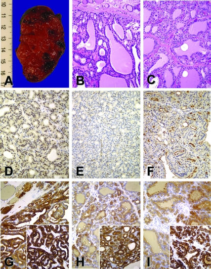
Figure 3.
A, Gross appearance of right thyroid lobe from the propositus of family B; B and C, hematoxylin-eosin stain of thyroid tissue from the propositi of family A (B) and family B (C); D and E, lack of apical membrane staining for pendrin with PS1Ab, an antibody against the first 15 amino acids of human pendrin, with perinuclear enhanced halo in thyroid follicle cells from the propositi of family A (D) and weak cytoplasmic staining in family B (E); F, strong apical membrane PS1Ab staining is seen in Graves’ disease; G–I, Tg and TPO immunoreactivity (inset) was found in both propositi and control thyroid glands: the proposita of family A (G), the propositus of family B (H), and Graves’ disease (I). All original magnifications, ×200.

