
Figure 4.
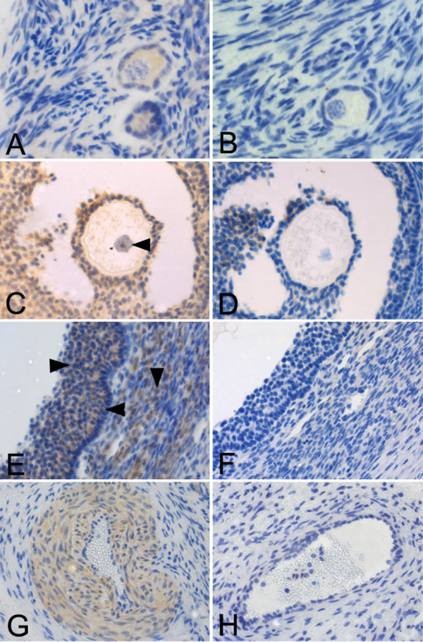
Ovarian localization of MYST4 protein by immunohistochemistry. Ovarian sections showing: primordial follicles (A, B); antral stage follicle, arrowhead showing oocyte nuclei (C, D); portion of large antral follicles, arrowheads indicating granulosa cells (right), theca (down) and basal lamina (left) (E, F) and blood vessels (G, H). Positive sections were incubated with anti-MYST4 (A, C, E, G,) and negatives were prepared by peptide-blocking assay (B, D, F, H). Original magnifications: 200× (C, D, E, F, G, H) and 400× (A, B).