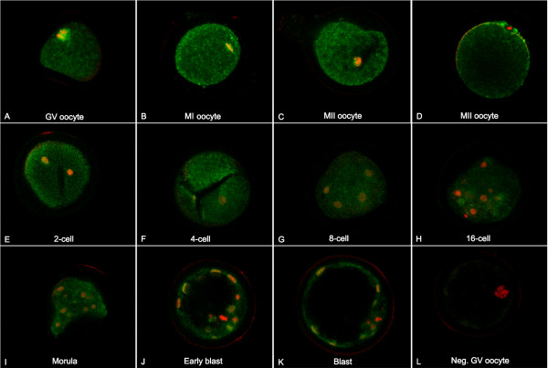
Figure 6.
Localization of MYST4 in oocyte and early embryo development. Confocal representation of oocytes (GV, MI and MII) or embryos (2-, 4-, 8-, 16-cell, morula, early blastocyst and blastocyst) stained with an anti-MYST4 antibody (green signal) and with propidium iodide (red signal) to visualize the DNA. Original magnification 600×.

