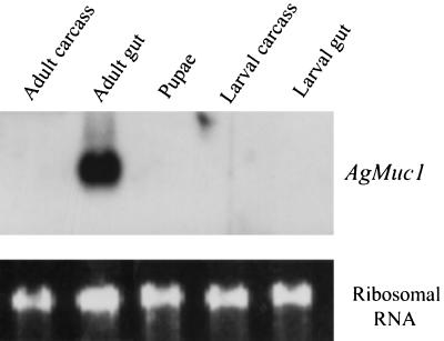
Figure 2.
Developmental and tissue specificity of AgMuc1 expression. (Upper) Autoradiogram of a Northern blot of RNAs (≈5 μg per lane) isolated from the indicated tissues. The blot was hybridized overnight with a 32P-labeled AgMuc1 cDNA probe, washed, and exposed to film for 4 h. (Lower) Staining of ribosomal RNA with ethidium bromide to indicate the amount of RNA analyzed in each lane.

