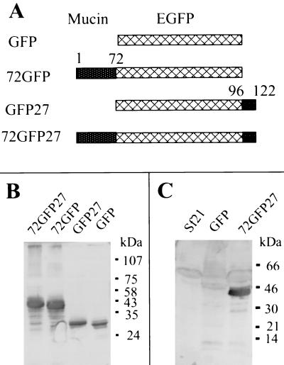
Figure 4.
Expression and characterization of mucin-GFP fusion proteins encoded by recombinant baculoviruses. (A) Diagrams showing recombinant protein structures. Lattice-patterned rectangles, GFP reporter protein; stippled rectangles, AgMuc1 sequences (see Fig. 1A). (B) Western blotting analysis of baculovirus-expressed GFP and GFP-mucin fusion proteins with an anti-GFP antibody. Total proteins from cells infected with different recombinant baculoviruses were loaded on each lane. A polyclonal antibody against GFP was used to probe the Western blots. The structure of the recombinant proteins indicated at the top of each lane is given in A. (C) Western blotting analysis of the baculovirus-expressed mucin-GFP protein with antiserum against total midgut microvilli. The Sf21 lane contains control cell lysate. Migration of marker proteins is indicated on the right side of B and C.

