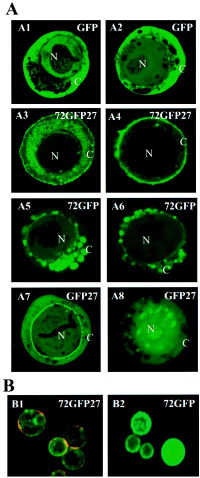
Figure 5.
Localization of baculovirus-expressed GFP fusion proteins in Sf21 cells. (A) Sf21 cells were infected with recombinant baculoviruses that express the GFP, 72GFP27, GFP27, and 72GFP recombinant constructs (Fig. 4A) under the control of the viral polyhedrin promoter. Representative images recorded at 30 h after infection (A1, A3, A5, and A7) and at 60 h after infection (A2, A4, A6, and A8) are shown. The green is due to fluorescence of GFP or of GFP fusion proteins. N, Nucleus; C, cytoplasm. (×1000.) (B) Immunological localization of mucin-GFP fusion proteins. Living Sf21 cells expressing fusion proteins 72GFP27 or 72GFP (as shown in A4 and A6, respectively) were incubated with a rabbit anti-GFP antibody followed by incubation with a rhodamine-conjugated anti-rabbit IgG secondary antibody, thus detecting GFP-containing proteins only if exposed to the surface (red channel). The distribution of GFP, independent of its location in the cell, was detected by its inherent fluorescence (green channel). The images shown in B1 and B2 were collected while using identical settings for the green and red channels, and images from the red and green channels were then merged. (×250.)

