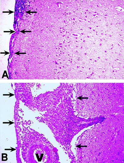
Figure 5.
Histopathology of meninges and brain of infected rabbits (21 days). (A) Rabbit infected with BCG Montreal (v). Mild focal inflammatory response is seen within the meninges (arrows). There is no evidence of perivascular tissue damage or leukocytic infiltration within the neuropil. (B) Rabbit infected with BCG mTNF-α. Marked thickening of the leptomeninges and distension of the arachnoid space by large numbers of inflammatory cells are observed (arrows). Note extension of perivascular inflammation to neuropil. Sections are stained with hematoxylin and eosin. Magnification ×25. Blood vessels are labeled V.

