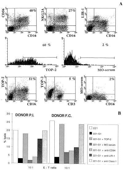
Figure 4.
Surface expression of CD94/NKG2A, LIR-1 and p49 receptors in lymphoid cells freshly isolated from placenta at term and their role in the HLA-G1 recognition. The lymphoid population analyzed was freshly derived from maternal decidua tissues of placenta at term. (A) Cells were stained with the indicated combinations of antibodies of different isotype, followed by appropriate isotype-specific second reagents, and were analyzed by double fluorescence analysis (Upper). The TOP-2 mouse antiserum (Lower) specifically recognizes the p49 receptor, while a control mouse serum (MO-serum) represents a control serum derived from a nonimmunized mouse (x axis, green fluorescence; y axis, red fluorescence). In this case, an anti-mouse IgG1 (PE-conjugated) has been used as second reagent. Note that, in double fluorescence experiments, both anti-CD16 and anti-CD3 mAbs belonged to the IgG2a subclass. (B) Lymphoid populations, freshly isolated from two placentas at term, were analyzed for the ability to lyse 221 cells transfected or not with HLA-G1. The cytolytic activity against 221-G1 was also assessed in the presence of Y9 (anti-CD94) or M401 (anti-LIR-1) mAb, the anti-p49 TOP-2 antiserum, and a MO-serum. A6136 (IgM) mAb in combination with 6A4 F(ab′)2, both directed to HLA class I molecules and reacting with HLA-G1, were also used. Note that masking of p49 restored lysis to an extent similar to that obtained by masking HLA-G1. Each histogram represents the mean of triplicate experiments.

