
Figure 2.
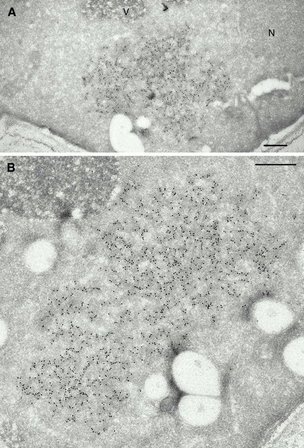
Cytoplasmic aggregates in [URE3] cells are labeled by anti-Ure2p antibodies. Cryosections were incubated with an affinity-purified antibody against the COOH-terminal domain of Ure2p. 10 nm gold particles show highly specific binding of the antibodies to cytoplasmic aggregates (A and B). Note the almost complete absence of label elsewhere in the cell. In the aggregates, gold particles are often locally organized in linear strands, suggesting multiple labeling of filaments. N, nucleus; V, vacuole. Bar, 0.5 μm.