
Figure 6.
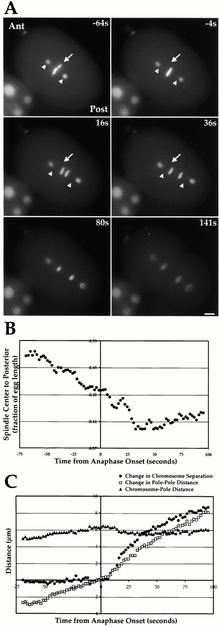
Polarity-cued forces begin moving the spindle towards the embryo posterior before anaphase onset. (A) Stills from a video of a C. elegans embryo expressing GFP-histone to mark the DNA and GFP–γ-tubulin to mark the spindle poles. The embryo anterior (Ant) is on the top left and the embryo posterior (Post) on the lower right. Time in seconds on the upper right of each panel is relative to anaphase onset. For reference, the position of the spindle center (arrow) and the two spindle poles (arrowheads) at the first time point (−64s) are marked on the subsequent three panels. (B) The distance between the spindle center and the embryo posterior (expressed as a fraction of egg length) is plotted. About half of the posterior movement of the spindle occurs before anaphase onset. (C) Chromosome segregation during the first mitotic division of the C. elegans embryo is primarily due to anaphase B. Chromosome-to-pole distance (black triangles), change in chromosome separation (medium gray circles), and change in pole-to-pole distance (light grey squares) are plotted relative to anaphase onset. Chromosome separation is the distance between the polar edges of the separating chromosomes measured along the pole–pole axis. Changes in chromosome separation and pole-to-pole distance were obtained by subtracting the relevant values 4 s before anaphase onset. Online supplemental videos are available at http://www.jcb.org/cgi/content/full/153/6/1209/DC1. Bar, 5 μm.