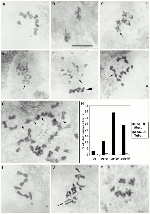
Figure 1.

Mitotic figures from wild-type, polo 9, and polo 10 brains. (A) A wild-type metaphase figure. (B) A wild-type anaphase figure. (C) Hypercondensed mitotic chromosomes from polo 9 . The arrow points to a pair of separated sister chromatids. (D) polo 10 cell showing separated sisters (arrow) that could be at early anaphase. (E) An anaphase (arrow) and metaphase (arrowhead) figure from polo 10 . (F) A tetraploid polo 10 cell in which several sister chromatids appear separated throughout their length, and yet joined at their telomeres (arrow). (G) A polyploid polo 9 cell in which chromosomes form long chains attached at their telomeres (arrows). (H) Bar graph showing the proportion of cells at prophase and metaphase (Pro. & Met.) or anaphase and telophase (Ana. & Tel.) in wild-type (wt), polo, polo 9, and polo 10 brains. (I) Wild-type cell from a larval brain treated for 4 h with colchicine. (J) polo 10 cell after 30 min colchicine treatment. The arrow marks separated sisters. (K) polo 10 cell after 2 h colchicine treatment.
