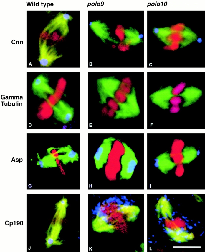
Figure 3.

Distribution of centrosomal antigens in wild-type, polo 9, and polo 10 cells. In all cases, wild-type cells are shown in the left panels, polo 9 cells the middle panels, and polo 10 cells the right panels. Spindle microtubules revealed by immunostaining with the YL1/2 antibody are shown in green, DNA stained with propidium iodide in red, and the centrosomal antigen in blue. (A–C) Centrosomin (Cnn) is revealed using a rabbit antibody from Heuer et al. 1995. (D–F) γ-Tubulin was detected using the mouse monoclonal antibody GTU88 (Sigma-Aldrich). (G–I) The Asp was detected using the rabbit antibody Rb3133 (Saunders et al. 1997). (J–L) CP190 was detected using the rabbit antibody RB188 (Whitfield et al. 1988). Bar, 5 μm.
