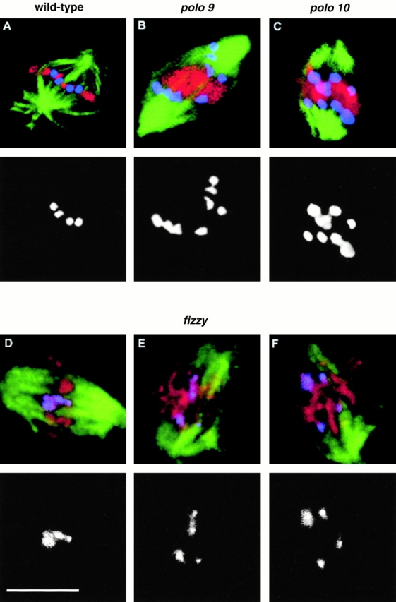
Figure 4.

Localization of Prod in wild-type (A), polo (B and C), and fizzy (D–F) cells. Spindle microtubules stained with the rat monoclonal antibody YL1/2 are stained green. DNA is stained red. Prod (blue) was detected using a rabbit antibody (Torok et al. 1997). Merged images are shown in the top panels with the separated channel for Prod staining below. Bar, 5 μm. 79.3% of 213 clear pairs of sister centromeric regions were scored as having separated in immunostained polo 9 cells. A similar frequency of separation (78.6% of 112 pairs) was observed in polo 10 cells. Centromere separation was not observed by anti-Prod staining in the fizzy x4 mutant.
