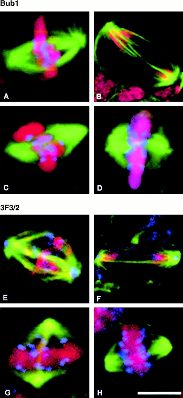
Figure 5.

Association of Bub1 and the 3F3/2 epitope with the mitotic apparatus in wild-type and polo mutant cells. In all panels, spindle microtubules are stained green and DNA stained red. Bub1 (A–F) or 3F3/2 (G–L) staining are shown in blue. (A and B) Wild-type cells at metaphase and anaphase, respectively. (C and D) polo 9 cells showing Bub1 staining with the rabbit antibody Rb666 (gift of C. Sunkel, University of Porto, Porto, Portugal) on the separated kinetochores. (E and F) Wild-type cells at metaphase and anaphase, respectively, stained with the 3F3/2 mouse monoclonal antibody (gift of G. Gorbsky, University of Oklahoma, Oklahoma City, Oklahoma). Note the presence of the 3F3/2 epitope at centrosomes. (G and H) polo 10 cells showing 3F3/2 staining on the separated kinetochores, and absent from the spindle poles. Bar, 5 μm.
