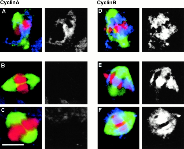
Figure 6.
Cyclins A and B in wild-type and polo mutant cells. In all panels, spindle microtubules are green, DNA red, and cyclin blue in the merged images. The monochromatic image is of cyclin staining alone using either the antibody Rb270 to detect cyclin A or Rb271 to detect cyclin B (Whitfield et al. 1990). (A) Cyclin A in a wild-type cell at prometaphase. (B and C) Absence of cyclin A staining in polo 9 and polo 10 mutant cells respectively. (D) Cyclin B staining in a wild-type cell at metaphase. (E and F) Cyclin B staining of polo 9 and polo 10 cells, respectively. Bar, 5 μm.

