During myogenesis, proliferating myoblasts withdraw from the cell cycle and fuse to form an ordered array of large, multinucleated muscle fibers. This highly regulated process can be divided temporally into a series of complex steps: commitment to a myoblast phenotype, acquisition of fusion competence, recognition and adhesion of like myoblasts, fusion of myoblasts into multinucleated myotubes, and differentiation of myotubes into muscle fibers. The end result is differentiated muscle fibers that contain several characteristic proteins, including: specific types of actin, myosin, tropomyosin, and troponin (which form part of the contractile apparatus), creatine phosphokinase, nicotinic receptors to respond to acetylcholine released from motor nerves, and various types of voltage-gated ion channels to generate actions potentials.
Commitment to a myoblast phenotype is an event that occurs early during embryonic development (Cossu et al. 1996). The steps that occur down stream of this commitment, however, are still being worked out. Like many developmental processes, myogenesis is complex and difficult to investigate in normal vertebrate embryos. Fortunately, crucial steps in myogenesis can be recapitulated in tissue culture. This discovery, made several years ago, has greatly facilitated research in this area. Using myoblast fusion as a relatively easy readout of differentiation, many researchers have investigated molecular mechanisms involved in aspects of myogenesis. A central focus of this research has been to identify molecules that promote myoblast fusion and to determine changes in these cells as they differentiate. Several essential molecules involved in fusion have been identified, including cell adhesion molecules, calcium and calmodulin, metalloproteases, phospholipases, and lipids (Yagami-Hiromasa et al. 1995; Tachibana and Hemler 1999).
As an alternative to tissue culture, some groups are working with Drosophila, thus enabling them to identify molecules crucial to myogenesis using a genetic approach. Myogenesis in Drosophila is similar to that in vertebrates (Doberstein et al. 1997; Frasch 1999), and so it is likely that molecules that are important for myoblast differentiation and fusion in Drosophila will have homologues in vertebrates. A number of Drosophila genes that appear to be involved in myogenesis include: rolling stone (rost), myoblast city (mbc), blown fuse (blow), dumbfounded (duf), and stick-and-stones (sns) (Frasch and Leptin 2000). Recently, research has shown that the gene products of duf and sns are novel members of the immunoglobulin superfamily differentially expressed on developing myoblasts (Bour et al. 2000; Ruiz-Gomez et al. 2000). Both molecules act during the early stages of myoblast fusion and null mutations in either the duf gene or the sns gene prevent myoblasts from fusing.
Although cell-adhesion molecules are clearly necessary for myoblast fusion, they are not sufficient. The work of Bernheim and colleagues, including their interesting report in this issue (Fischer-Lougheed et al. 2001), indicates that human myoblasts must also express particular types of ion channels. A brief review of some of this work will help explain why these ion channels are necessary in myoblast fusion.
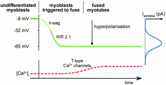
Concomitant with fusion, the electrical potential across the cell membrane of perfusion myoblasts hyperpolarizes from −10 to approximately −65 mV in newly formed myotubes, a potential required by most voltage-gated ion channels to function effectively. This hyperpolarization is achieved in two steps (Fig. 1, green line). First, fusion-competent myoblasts express a noninactivating, voltage-gated potassium channel, referred to as ether-a-go-go, or h-eag (Bernheim et al. 1996). These channels open at depolarized membrane potentials and close when the membrane potential hyperpolarizes. The result of expressing h-eag channels is that the membrane potential of fusion-competent myoblasts hyperpolarizes to approximately −30 mV. Shortly thereafter, these myoblasts express a second potassium channel, referred to as an inward rectifier or KIR (Liu et al. 1998); these channels are open at hyperpolarized membrane potentials, but closed at depolarized potentials. Expressing these KIR channels hyperpolarizes the membrane potential from approximately −30 to −65 mV. To change the membrane potential from approximately −10 to −65 mV, h-eag and KIR channels must appear sequentially—if myoblasts express h-eag channels only, then the membrane potential hyperpolarizes approximately −30 mV, and if myoblasts express KIR channels without h-eag channels, then the membrane potential remains at approximately −10 mV because KIR channels generate very little current at this potential and therefore cannot hyperpolarize the membrane.
Figure 1.
Model of events leading to an increase in intracellular calcium in fusing myoblasts (adapted from Bijlenga et al. 2000). The green line represents the change in the membrane potential over time. The membrane potential is approximately −8 mV in undifferentiated myoblasts (dark green). When myoblasts are rigged to fuse, they express h-eag, which brings the membrane potential to approximately −30 mV (light green); then the myoblasts express KIR2.1, which brings the membrane potential to approximately −65 mV (green). Shown at the right in blue is the window current for the T-type calcium channels as a function of membrane potential. At approximately −65 mV, the window current is maximum. The bottom curve in red shows the increase in intracellular calcium, which promotes fusion.
What does this have to do with myoblast fusion? Interestingly, Fischer-Lougheed et al. 2001 demonstrate that blocking the expression of KIR channels in fusion-competent myoblast prevents fusion. To show this, they had to determine which type of KIR channel was expressed in these myoblasts, as several KIR genes are known to exist. In their study, Fischer-Lougheed et al. 2001 used routine electrophysiological and molecular biological techniques to identify the channel as KIR2.1 inward rectifier. Next, they expressed antisense constructs for KIR2.1 and showed that decreasing KIR2.1 prevented these myoblasts from hyperpolarization to approximately −65 mV; instead, the membrane potential only hyperpolarized to approximately −30 mV, indicating that expression of functional h-eag channels was unaffected by the decrease in KIR. More to the point, decreasing KIR2.1 expression prevented these myoblasts from fusing.
The question is, why are KIR2.1 channels essential for fusion? By themselves, the extracellular portions of KIR2.1 are not known to have cell-adhesion properties. More likely, fusion occurs because the membrane hyperpolarizes, allowing calcium to enter the cell. It has been known for some time that extracellular calcium is required for fusion (Wakelam 1985). Moreover, a rise in intracellular calcium precedes fusion (Entwistle et al. 1988; Rapuano et al. 1989) (Fig. 1, red line). Bijlenga et al. 2000 propose that this rise in intracellular calcium results from calcium entry through particular voltage-gated calcium channels, referred to as T-type, that are expressed on pre-fusion myoblasts. At hyperpolarized membrane potentials, these T-type channels are closed; when the membrane depolarizes, these channels open transiently, and then inactivate, or close. To remove this inactivation, the membrane potential must hyperpolarize. It turns out that at a critical membrane potential, one that is sufficiently depolarized to open channels but sufficiently hyperpolarized to remove inactivation, T-type calcium channels continuously cycle from closed to open to inactivate and back again. At this membrane potential, a small proportion of these channels will always be open and allow calcium into the myoblast; this small inflow of calcium ions is called the “window current” (Fig. 1, blue line). Bijlenga et al. 2000 propose that when myoblasts express KIR2.1 and hyperpolarize to approximately −65 mV, this membrane potential activates the window current for T-type calcium channels (Fig. 1). The resulting calcium influx triggers fusion.
These intriguing findings raise a number of questions: What happens downstream of this calcium signal to promote fusion? Does this increase in intracellular calcium activate calcium-dependent kinases and phosphatases and perhaps promote fusion by signaling through cell-adhesion molecules? Are T-type calcium channels the only pathway for calcium to enter the myoblast (see, for example, Entwistle et al. 1988; Rapuano et al. 1989)? How does myogenesis proceed in mice with targeted mutations in the KIR2.1 gene?
Acknowledgments
I am grateful to P. Holland for discussions and to L. Cooper for suggestions on the manuscript.
This work was supported by the Canadian Institutes for Health Research.
References
- Bernheim L., Lui J.-H., Espinos E., Haenggeli C.A., Fisher-Lougheed J., Bader C.R. Contribution of a non-inactivating potassium current to the resting potential of fusion-competent human myoblasts. J. Physiol. 1996;493:129–141. doi: 10.1113/jphysiol.1996.sp021369. [DOI] [PMC free article] [PubMed] [Google Scholar]
- Bijlenga P., Liu J.-H., Espinos E., Haenggeli C.A., Fischer-Lougheed J., Bader C.R., Bernheim L. T-type alpha 1H Ca2+ channels are involved in Ca2+ signaling during terminal differentiation (fusion) of human myoblasts. Proc. Natl. Acad. Sci. USA. 2000;97:7627–7632. doi: 10.1073/pnas.97.13.7627. [DOI] [PMC free article] [PubMed] [Google Scholar]
- Bour B.A., Chakravarti M., West J.M., Abmayr S.M. Drosophila SNS, a member of the immunoglobulin superfamily that is essential for myoblast fusion. Genes Dev. 2000;14:1498–1511. [PMC free article] [PubMed] [Google Scholar]
- Cossu E.N., Tajbakhsh S., Buckingham M. How is myogenesis initiated in the embryo? Trends Genet. 1996;12:218–223. doi: 10.1016/0168-9525(96)10025-1. [DOI] [PubMed] [Google Scholar]
- Doberstein S.K., Fetter R.D., Mehta A.Y., Goodman C.S. Genetic analysis of myoblast fusionblown fuse is required for progression beyond the pre-fusion complex. J. Cell Biol. 1997;136:1249–1261. doi: 10.1083/jcb.136.6.1249. [DOI] [PMC free article] [PubMed] [Google Scholar]
- Entwistle A., Zalin R.J., Bevan S., Warner A.E. The control of chick myoblast fusion by ion channels operated by prostaglandins and acetylcholine. J. Cell Biol. 1988;106:1693–1702. doi: 10.1083/jcb.106.5.1693. [DOI] [PMC free article] [PubMed] [Google Scholar]
- Fischer-Lougheed J., Liu J.-H., Espinos E., Mordasini D., Bader C.R., Belin D., Bernheim L. Human myoblast fusion requires expression of functional inward rectifier Kir2.1 channels. J. Cell Biol. 2001;153:677–685. doi: 10.1083/jcb.153.4.677. [DOI] [PMC free article] [PubMed] [Google Scholar]
- Frasch M. Controls in patterning and diversification of somatic muscles during Drosophila embryogenesis. Curr. Opin. Genet. Dev. 1999;9:522–529. doi: 10.1016/s0959-437x(99)00014-3. [DOI] [PubMed] [Google Scholar]
- Frasch M., Leptin M. Mergers and acquisitionsunequal partnerships in Drosophila myoblast fusion. Cell. 2000;102:127–129. doi: 10.1016/s0092-8674(00)00016-7. [DOI] [PubMed] [Google Scholar]
- Liu J.-H., Bijlenga P., Fischer-Lougheed J., Occhiodoro T., Kaelin A., Bader C.R., Bernheim L. Role of an inward rectifier K+ current and of hyperpolarization in human myoblast fusion. J. Physiol. 1998;510:467–476. doi: 10.1111/j.1469-7793.1998.467bk.x. [DOI] [PMC free article] [PubMed] [Google Scholar]
- Rapuano M., Ross A.F., Prives J. Opposing effects of calcium entry and phorbol esters on fusion of chick muscle cells. Dev. Biol. 1989;134:271–278. doi: 10.1016/0012-1606(89)90099-7. [DOI] [PubMed] [Google Scholar]
- Ruiz-Gomez M., Coutts N., Price A., Taylor M.V., Bates M. Drosophila dumbfoundeda myoblast attractant essential for fusion. Cell. 2000;102:189–198. doi: 10.1016/s0092-8674(00)00024-6. [DOI] [PubMed] [Google Scholar]
- Tachibana I., Hemler M.E. Role of transmembrane 4 superfamily (TM4SF) proteins CD9 and CD81 in muscle cell fusion and myotube maintenance. J. Cell Biol. 1999;146:893–904. doi: 10.1083/jcb.146.4.893. [DOI] [PMC free article] [PubMed] [Google Scholar]
- Wakelam M.J.O. The fusion of myoblasts. Biochem. J. 1985;228:1–12. doi: 10.1042/bj2280001. [DOI] [PMC free article] [PubMed] [Google Scholar]
- Yagami-Hiromasa T., Sato T., Kurisaki T., Kamijo K., Nabeshima Y., Fujisawa-Sehara A. A metalloprotease-disintegrin participating in myoblast fusion. Nature. 1995;377:652–656. doi: 10.1038/377652a0. [DOI] [PubMed] [Google Scholar]


