
Figure 2.

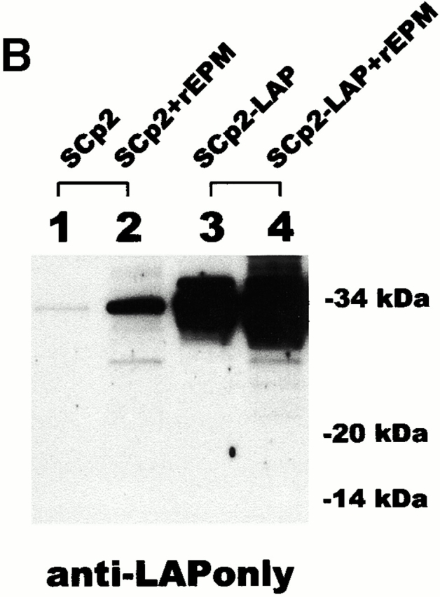
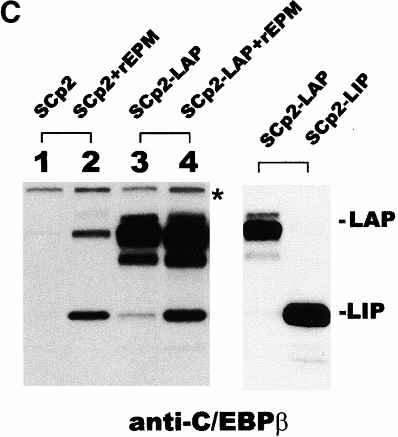
Minimal proteolysis of LAP occurs during sample preparation. (A) Diagram depicting the targeted location of the anti-LAPonly antibody and of the commercial anti-C/EBPβ antibody. (B) Western blot probed with the anti-LAPonly antibody. (C) Western blots probed with commercial anti-C/EBPβ antibody. Left: Parallel blot to B; asterisk, cross-reactive material. Right: Blot of SCp2 cells transiently transfected with LAP or LIP expression plasmids. Results shown are typical of two independent experiments.