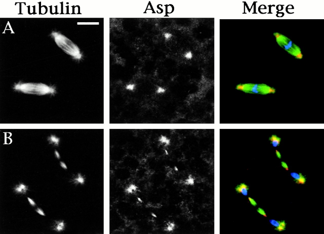
Figure 2.
Asp localization in cells of wild-type Drosophila embryos. In the merged images, DNA is colored in blue, tubulin in green, and Asp in orange. (A) Metaphases showing Asp localization at the spindle poles; (B) telophases showing Asp accumulation at the minus ends of central spindle microtubules. Bar, 10 μm.

