
Figure 3.
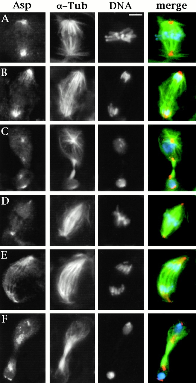
Asp localization in neuroblasts of wild-type and asl larval brains. Cells were stained for α-tubulin, Asp, and DNA (by DAPI). In the merged images, DNA is colored in blue, tubulin in green, and Asp in orange. (A–C) Wild-type neuroblasts; (D–F) asl neuroblasts; (A and D) metaphases; (B and E) anaphases; (C and F) telophases. Note that both wild-type and asl neuroblasts divide asymmetrically, giving rise to two daughter cells of different sizes (C and F). For details on unequal neuroblast division see Giansanti et al. 2001. In metaphase and anaphase figures, Asp is localized at the spindle poles, wheras in telophases (C and F) it accumulates at both the poles and the extremities of the central spindle. Note that wild-type and asl neuroblasts exhibit similar patterns of Asp accumulation. Bar, 5 μm.