
Figure 4.
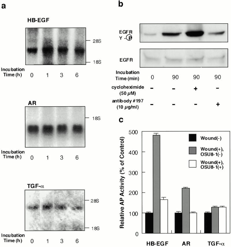
Characterization of EGFR-ligand shedding in keratinocytes. (a) Gene expression of EGFR ligands in wounded keratinocytes. After incubation of keratinocytes for the indicated time after wounding, total RNA was extracted and analyzed by Northern blotting. Sequential hybridization using the same membrane is shown. HB-EGF (2.4 kb), AR (1.4 kb), and TGF-α (4.8 kb) messages were detected. (b) Effect of cycloheximide and HB-EGF neutralizing antibody No. 197 on wound-induced EGFR activation in cultured keratinocytes. Keratinocytes were stimulated by tip-scraping in the presence or absence of cycloheximide (50 μM) or HB-EGF neutralizing antibody No. 197 (10 μg/ml). After incubating for the indicated times, the cells were lysed and subjected to immunoprecipitation with EGFR antibodies, fractionation by SDS-PAGE, and Western blotting. Half of the immunocomplex was used for the detection of tyrosine phosphorylation with phosphotyrosine antibody PY-20, and the other half for detection of EGFR protein with EGFR antibody. Cycloheximide (50 μM) did not abrogate the wound-induced tyrosine phosphorylation of EGFR. On the other hand, antibody No. 197 (10 μg/ml) suppressed wound-induced tyrosine phosphorylation of EGFR by 70%. (c) Quantitative analyses of EGFR ligand shedding. Stable transfectants of HaCat cells expressing nearly equal amounts of AP-tag HB-EGF, AP-tag AR, or AP-Tag TGF-α were incubated with or without 1 μM OSU8-1 for 60 min after wounding. AP activities in the conditioned media were measured. Each bar is the average of triplicate values.