Figure 5.
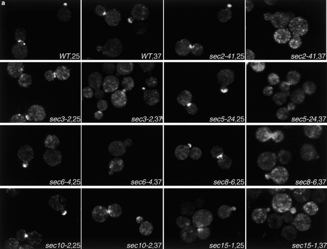
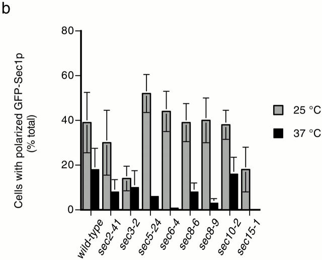
Localization of GFP-Sec1p in sec mutants. (a) Fluorescent images of GFP-Sec1p localization in wild-type (SEC+, NY1696), sec2-41 (NY2222), sec3-2 (NY2214), sec 5-24 (NY2215) sec6-4 (NY2216), sec8-6 (NY2217), sec8-9 (NY2218), sec10-2 (NY2219), and sec15-1 (NY2220) cells at 25°C and after a 10-min incubation at 37°C. Cells were fixed and viewed by epifluorescence microscopy. (b) Quantitation of GFP-Sec1p localization in wild-type and sec mutant cells. The average of duplicate experiments (except for wild-type, which was done in triplicate) is followed by a range that reflects the variability between experiments performed on different days. At 25°C, the percentage of cells with polarized GFP-Sec1 was as follows: wild-type, 39 ± 13% (n = 826); sec2-41, 30 ± 14% (n = 569); sec3-2, 14 ± 6% (n = 753); sec5-24, 52 ± 9% (n = 409); sec6-4, 44 ± 9% (n = 232); sec8-6, 39 ± 9% (n = 708); sec8-9, 40 ± 10% (n = 410); sec10-2, 38 ± 7% (n = 396); and sec15-1, 18 ± 10% (n = 351). At 37°C, the percentage of cells with polarized GFP-Sec1 was as follows: SEC+, 18 ± 5% (n = 1,046); sec2-41, 8 ± 7% (n = 722); sec3-2, 10 ± 8% (n = 945); sec5-24, 6 ± 0.15% (n = 462); sec6-4, 0.8 ± 0.25% (n = 403); sec8-6, 8 ± 4% (n = 816); sec8-9, 3 ± 2% (n = 419); sec10-2, 16 ± 8% (n = 307); and sec15-1, 0 (n = 554).


