
Figure 3.
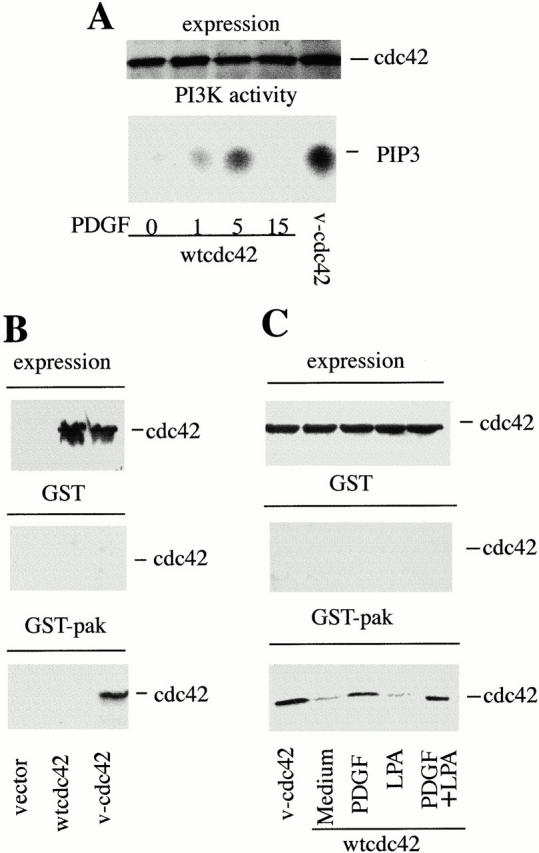
Stimulation of fibroblasts with PDGF activates Cdc42. 1.7 × 10⁶ NIH-3T3 cells were transfected with 10 μg of control cDNA, cDNA encoding myc-wt-Cdc42, or myc-V12-Cdc42 (myc-v-Cdc42). After transfection, cells were incubated as in Fig. 2. In A, PDGF-R stimulation (50 ng/ml) was performed for the indicated periods of time (in minutes). (A) Lysates were resolved in 12% SDS-PAGE (60 μg, lane), transferred to nitrocellulose, and membranes were blotted with anti-myc Ab (top). Alternatively, lysates were immunoprecipitated (300 μg/sample) using anti-myc Ab, and the PI3K activity associated with myc-Cdc42 was measured in vitro (bottom). (B) Cdc42 expression in the transfected samples (indicated) was analyzed as in A, or, alternatively, lysates (300 μg/sample) were passed through a GST-glutathione-Sepharose column (control) or a Pak-1-GST-glutathione-Sepharose column. The Myc-Cdc42 bound to the columns was examined by Western blot using anti-myc Ab. (C) Cells were transfected, starved, and stimulated for 5 min with PDGF and LPA (doses as in Fig. 2 C). Cell lysates were processed as in B. Specific wt-Cdc42 binding to Pak-1 columns is seen in PDGF-stimulated cells. The figure illustrates one representative experiment of three performed with similar results.