
Figure 7.
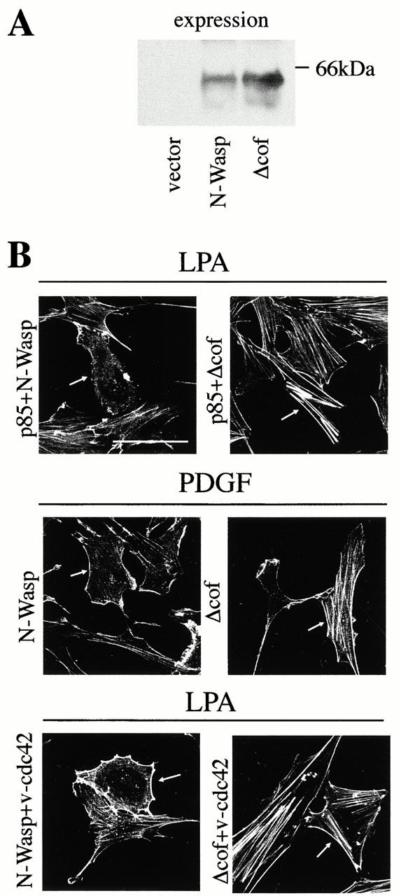
N-WASP mediates the inhibition of actin stress fibers induced by PDGF and by p85α. 2 × 10⁵ NIH-3T3 cells were transfected as in Fig. 2 with a total of 5 μg cDNA. Different combinations of vectors encoding HA-p85α, N-WASP, Δ-cof-N-WASP, and myc-v-Cdc42 were used (indicated). (A) 50 μg of lysates from cells transfected with cDNAs encoding control vector, or N-WASP, or Δ-cof-N-WASP were resolved by SDS-PAGE, transferred onto nitrocellulose, and examined by Western blot using anti-N-WASP Ab. (B) After transfection with the indicated plasmids, cells were cultured and activated as in Fig. 2. Cells were subsequently fixed and stained simultaneously with FITC-phalloidin (depicted) and with anti-myc, anti-HA, or anti–N-WASP Ab (indirect immunofluorescence, not shown) to detect positive transfected cells (indicated by an arrow). Throughout the experiment, Δ-cof-N-WASP expression blocked stress fiber disassembly, which was induced by p85α, PDGF, or v-Cdc42. The figure illustrates one representative experiment of five performed with similar results. Bar, 100 μm.