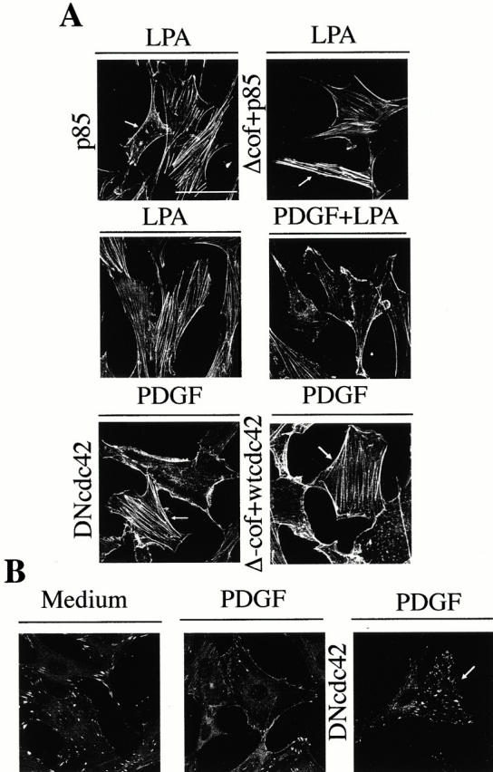
Figure 8.

Actin cytoskeleton changes induced by PDGF stimulation or p85α expression in NIH-3T3 cells cultured on collagen. NIH-3T3 cells were cultured on collagen VI–coated plates. The samples (2 × 10⁵ NIH-3T3 cells) were transfected with different combinations of vectors encoding HA-p85α, Δ-cof-N-WASP, myc-wt-Cdc42, and myc-DN-Cdc42 (indicated). After transfection, cells were incubated in complete medium, starved, and subsequently activated as in Fig. 2. (A) Cells were fixed and stained with FITC-phalloidin or, in the case of transfected cells stained simultaneously with FITC-phalloidin (depicted) and anti-myc, anti-HA, or anti-N-WASP Ab to detect transfected cells (indicated by an arrow). (B) Paxillin staining of NIH-3T3 cells cultured on collagen and transfected, starved, and treated as in A (indicated). The figure shows a representative experiment of four performed with similar results. Bar, 100 μm.
