
Figure 7.
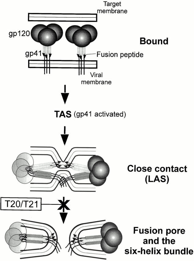
A proposed sequence of events for Env-mediated membrane fusion. The relative positions of HR1 and HR2 in the bound state are not known and only HR1 is depicted. (Neither CD4 nor the coreceptors are shown.) When gp41 is activated, the grooves of the central coiled coil (light gray bars with fusion peptide indicated by arrows) and the COOH-terminal helices (dark gray bars attached to the TM domain and cytoplasmic tail) have become exposed. The gp120 on the left is made transparent, for clarity. When gp41 further reconfigures into a six-helix bundle, the fusion peptide and TM domain (in different membranes) are forced toward each other and induce pore formation (probably by rupturing the hemifusion diaphragm, not shown).