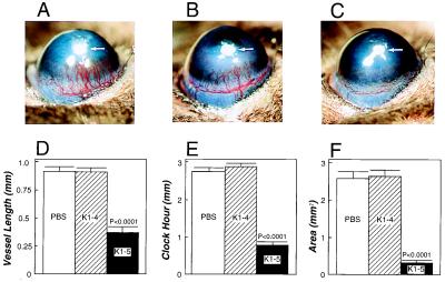
Figure 5.
Inhibition of mouse corneal neovascularization. Pellets containing sucrose aluminum sulfate, hydron, and 80 ng of FGF were implanted into corneal micropockets of mice. Corneas were photographed with a stereomicroscope on day 6 after FGF implantation and positions of implanted pellets were indicated by arrows in A–C. (A) Cornea of a control mouse receiving daily subcutaneous injection of PBS. (B) An example of the mouse cornea treated with daily subcutaneous injections of K1–4 (2 mg/kg). (C) An example of the mouse cornea treated with daily subcutaneous injections of K1–5 (2 mg/kg). Five mice of each treated and control group were used. (D) Maximal vessel length. (E) Clock hours of circumferential neovascularization. (F) Area of neovascularization. All data in D–F are presented as the mean ± SEM from 10 corneas in each group.

