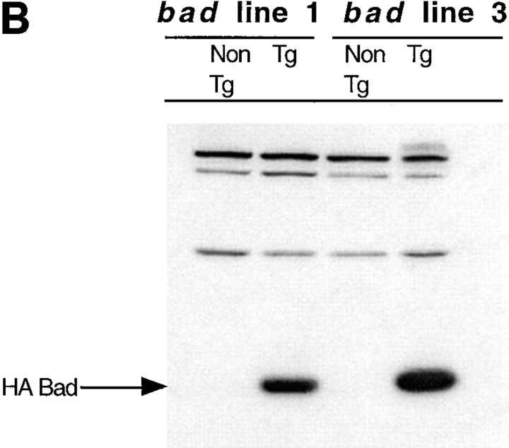
Figure 2.
bad transgene. (A) The mouse bad cDNA including a 5′ HA epitope was cloned into the EcoRI site of the human CD2 VA expression vector. The SalI-XbaI fragment was then isolated for microinjection. Western blot analysis of transgene expression in the two transgenic lines studied was carried out using total cell extract of thymocytes. Equal amounts of protein were loaded in each lane. (B) The blot was probed with the 12CA5 mAb against HA, to detect the presence of the transgene and (C) with a polyclonal anti-Bad antibody to compare the levels of endogenous and transgenic Bad expression.


