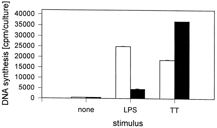
Figure 2.
Accessory activity of monocytes and dendritic cells during stimulation of T cells by LPS. Purified T lymphocytes (106/ml) were cultured in the presence of 10% freshly isolated autologous monocytes (white bars) or 10% autologous dendritic cells (black bars) and stimulated with LPS (S. friedenau, 1 μg/culture) in RPMI 1640 plus 10% HS in a final volume of 200 μl/culture. After 7 d of culture, cells were pulsed with [3H]TdR (0.2 μCi/culture), then harvested on glass filter mats, and the radioactivity was measured in a β-counter. The results of one of three experiments are given. Data are expressed as mean ± SD of three independent cultures.

