Figure 3.

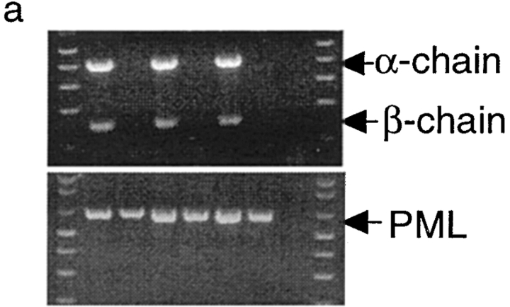
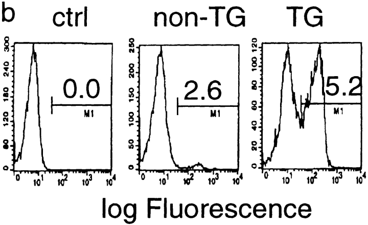
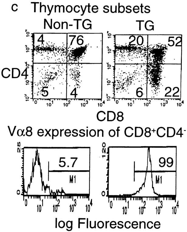
Endogenous expression of unmutated tumor antigen does not interfere with development of transgenic T cells expressing P1A-specific TCR. (a) Cointegration of TCR α- and β-transgene in transgenic founder mice. Tail DNA from 22 independent founder mice were screened by PCR for integration of transgenes, six of which are shown. Top, integration of transgenes into mouse tail genomic DNA. PCR reactions for the two chains were carried out separately, but PCR products from the same mice were pooled and analyzed in the same lane. Bottom, the quality of the genomic DNA is confirmed by similar amplification of exon 3 of the PML gene. (b) Expression of transgenic α chain among the TCR-transgenic (TCR-TG), nontransgenic (non-TG), or control PBL. PBL were either left unstained (ctrl) or stained with FITC–labeled anti-Vα8 mAb. (c) Development of transgenic T cells in BALB/c × TCR–TG F1 mice. Thymi from TCR-TG+ and TCR-TG− mice were analyzed by three-color flow cytometry using FITC–anti-Vα8, PE–anti-CD4, and cychrome–anti-CD8 mAbs. Data presented are expression of CD4 and CD8 coreceptors among total thymocytes (top) and expression of TCR α chain among CD8+CD4− T cells (bottom). (d) CD8 T cells in the spleen expressing transgenic TCR maintain naive phenotype. Spleen cells from TCR-TG mice were analyzed by three-color flow cytometry using FITC–anti-Vα8, PE–anti-CD62L, and cychrome–anti-CD8 mAbs. Data presented are histograms of PE channel among the CD8+ Vα8− (top) and CD8+Vα8+ (bottom) cells. Numbers presented in the panels are percentages of cells that fall within the gate indicated.


