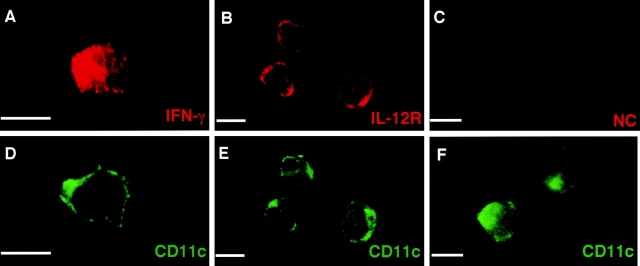
Figure 4.
Detection of intracellular IFN-γ in IL-12–stimulated DCs. Purified DCs were cultured in the presence of 1 ng IL-12 for 3 d. After surface staining with FITC-conjugated mAb against CD11c, cells were fixed and permeabilized. Cells were then incubated with rabbit polyclonal Ab against IFN-γ (A and B) and normal rabbit serum (E and F). Freshly isolated DCs were also incubated with rabbit polyclonal Ab against IL-12R (C and D). Samples were further stained with Rhodamine-conjugated goat anti-rabbit IgG. Bars, 10 μm.

