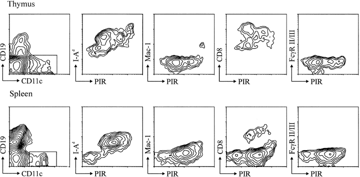
Figure 5.
PIR expression by thymic and splenic dendritic cells. (Top) Thy-1+ T and B220+ B cells were depleted from thymic cell suspensions by complement-mediated lysis, and the remaining cells were stained with the indicated mAbs. The CD11c+/CD19− cells, which comprised ∼1% of the initial MNC population, were analyzed for the expression of PIR and other cell surface markers (MHC I-Ad, Mac-1, CD8, and FcγRII/ III). (Bottom) Splenocytes were stained similarly, and the CD11c+/CD19− cells analyzed for other cell surface antigens.

