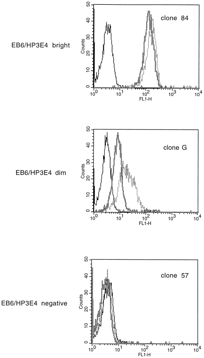
Figure 6.

Staining NK clones with mAbs to NK receptors reveals distinct phenotypes between clones that are sensitive to the transmembrane sequence of HLA-Cw6 and those that are not. Histograms of fluorescence intensity of NK clones stained with EB6 (thick gray line), HP3E4 (dotted line), and no first antibody (thin black line), followed by FITC- labeled goat anti–mouse Ig secondary antibody.
