Figure 8.
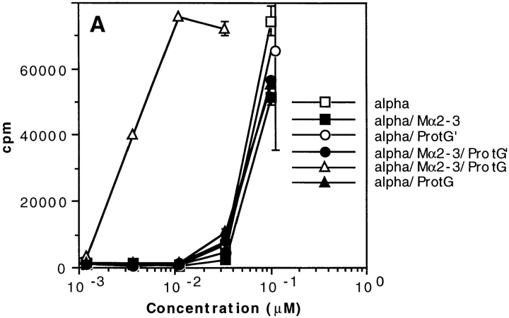
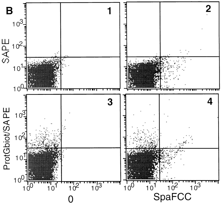
T cell presentation of toxin α in the presence of protein G from Streptococcus ssp. (A) Toxin α (alpha) was serially diluted and incubated overnight at 4°C in the absence or presence of fixed concentrations of mAb Mα2-3 (25 nM final) and protein G or protein G′ (0.1 μM final concentration for each derivative). 5 × 10⁵ splenocytes from BALB/c mice were then added to each well in the presence of 5 × 10⁴ T1B2. Cells were cultured for 24 h at 37°C, and IL-2 secretion was subsequently determined by CTLL assay. (B) Splenocytes (5 × 10⁵ cells) were incubated in the absence of IBP (B1) or presence of biotinylated protein G (protGbiot; B2), SpA–FCC (B3), or both (B4) for 30 min at 4°C. Cells were all incubated with SAPE and analyzed by flow cytometry.


