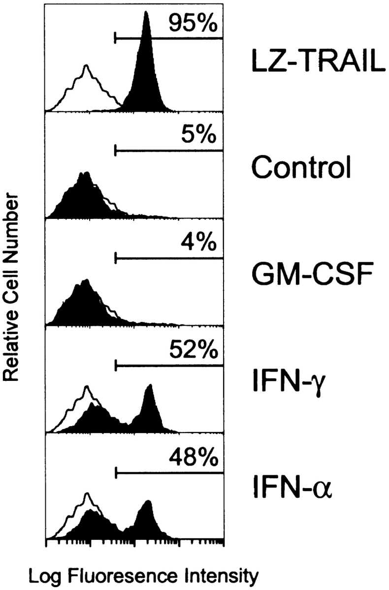
Figure 3.

Phosphatidylserine externalization on OVCAR3 tumor cells during apoptosis induced by IFN-stimulated, TRAIL- expressing Mφ. OVCAR3 tumor cells were cultured for 6 h in medium alone or in the presence of LZ-TRAIL (1 μg/ml), unstimulated, or cytokine (GM-CSF, IFN-γ, IFN-α [100 ng/ml for 12 h])–stimulated Mφ (E/T ratio 2:1). Cells were then stained with FITC–annexin V and analyzed by flow cytometry. The percent of FITC–annexin V positive tumor cells is indicated for each condition. Histograms represent 104 gated tumor cells. Similar results were seen with Mφ from three other donors.
