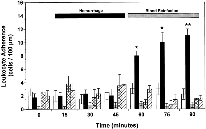
Figure 3.
Leukocyte adherence observed in peri-intestinal venules of wild-type mice, P-selectin–deficient (P-selectin−/−) mice, and wild-type mice given either anti–P-selectin mAb or rs.PSGL.Ig, and subjected to hemorrhagic shock. Bar heights represent means and brackets indicate ± SEM. *P < 0.05 and **P < 0.01 from control wild-type mice. White bars, control wild-type (n = 6); black bars, hemorrhage wild-type (n = 7); gray bars, hemorrhage P-selectin−/− (n = 6); cross-hatched bars, hemorrhage wild-type + anti–P-selectin mAb (n = 5); hatched bars, hemorrhage wild-type + rs.PSGL.Ig (n = 5).

