
Figure 4.
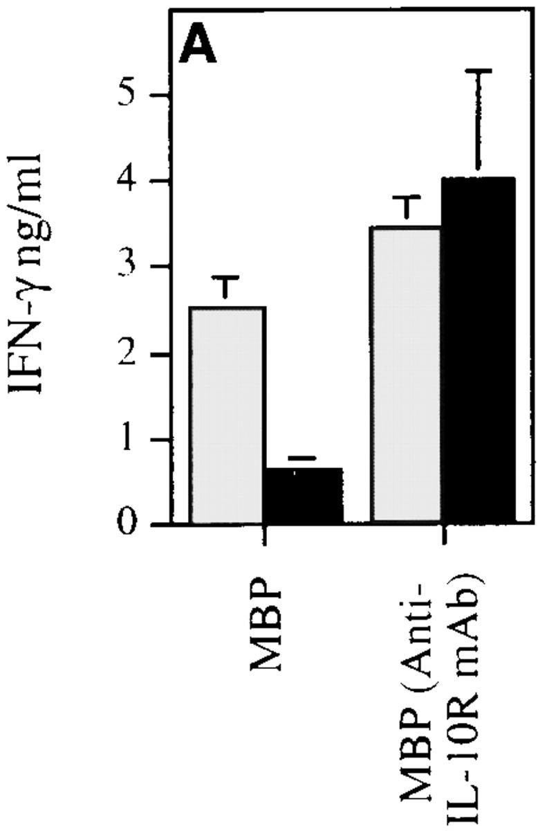
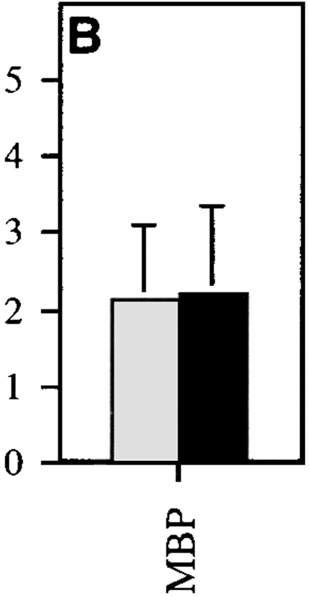
MBP-specific Th1 cells are induced in hIL-10Tg mice. hIL-10Tg mice (black bar) and control littermate mice (gray bar) were immunized twice at 1-wk intervals with MSCH emulsified in CFA. (A) 3 d after the last immunization, DLN cells from hIL-10Tg or control littermate mice were stimulated with MBP or MBP plus anti–IL-10 receptor mAb. One representative experiment out of three is shown. (B) CD4+ T cells were purified and stimulated in vitro with MBP plus irradiated T cell–depleted splenocytes from control CSJLF1/J mice for 60 h. The amount of IFN-γ in the supernatants was measured by ELISA. One representative experiment out of two is shown. The results are the mean of three or four individual mice/group ± SEM.