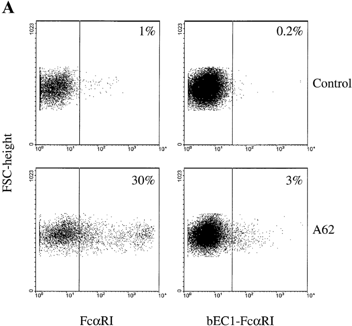
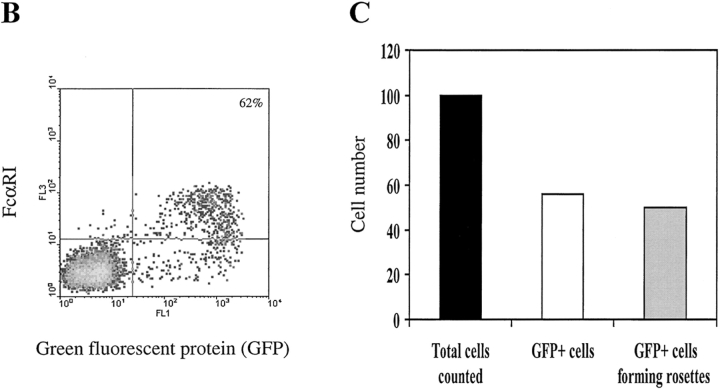
Figure 2.
Expression of chimeric FcR by COS-1 cells. (A) Cos-1 cells were transfected with the indicated constructs 2 d before harvesting and FACS® analysis. Cells were stained with either FcαRI EC2-specific mAb A62 (mouse IgG1) (bottom panels) or an appropriate isotype control (top panels), followed by a GAM IgG1 Tricolor reagent. Numbers in the top right corners of the plots refer to the percentage of positive cells. (B) Enrichment of FcαRI expression in COS-1 cells cotransfected with GFP. Cells transfected with both FcαRI and GFP were stained with FcαRI EC2-specific mAb A62 followed by GAM IgG1 Tricolor reagent and analyzed by FACS®. More than 60% of the GFP+ cells also express FcαRI. (C) Rosette formation by COS-1 cells cotransfected with both FcαRI and GFP and exposed to hIgA-coated beads. Rosettes were quantified as specified in Materials and Methods. Black bar, total number of cells counted; white bar, total number of GFP+ cells assessed by fluorescent microscopy; gray bar, number of GFP+ cells forming rosettes. Results shown are representative of three separate experiments.


