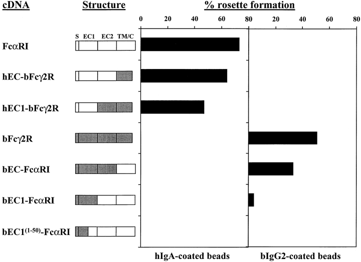
Figure 4.
Rosette formation by FcR/GFP cotransfected COS-1 cells. Schematic representation of wild-type and chimeric FcRs. Unshaded regions are derived from FcαRI, shaded regions from bFcγ2R. S, signal peptide; EC1, extracellular domain 1; EC2, extracellular domain 2; TM/C, transmembrane/cytoplasmic tail. GFP+ transfectants were purified from COS-1 cells cotransfected with GFP and FcR constructs as described. Ig-binding to FcRs coexpressed with GFP was assessed by rosetting with either hIgA- or bIgG2-coated beads. More than 200 cells were counted for each determination, and the number of cells binding four or more Ig-coated beads is expressed as percentage rosette formation. Results are representative of three separate experiments.

