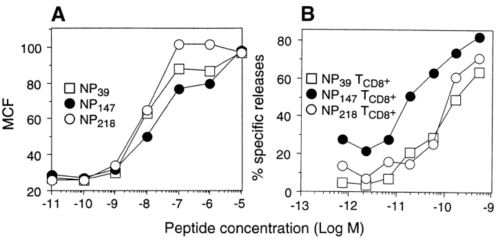
Figure 1.
Antigenicity of synthetic peptides corresponding to dominant and subdominant determinants. (A) T2-Kd cells were cultured for 14 h at 26°C and added to the wells containing synthetic peptides at the indicated concentrations. The samples were immediately shifted to 37°C and incubated for 2 h to denature Kd molecules lacking peptides and then stained with a fluorescein-conjugated anti-Kd mAb. The mean channel fluorescence (MCF) of viable cells was determined by flow cytometry. (B) Splenocytes from PR8-primed animals stimulated in vitro for 7 d with synthetic peptides corresponding to NP39–47, NP147–155, or NP218–226 were tested in a microcytotoxicity assay for their ability to lyse 51Cr- labeled P815 target cells incubated in I-10 with synthetic peptides at the indicated concentrations.

