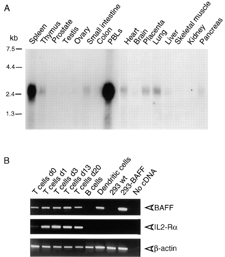
Figure 3.
Expression of BAFF. (A) Northern blots (2 μg poly A+ RNA per lane) of various human tissues were probed with BAFF antisense mRNA. (B) Reverse transcriptase amplification of BAFF, IL-2 receptor α chain (IL2-Rα), and actin from RNA of purified blood T cells at various time points of PHA activation, E-rosetting–negative blood cells (mostly B cells), in vitro–derived immature dendritic cells, 293 cells, and 293 cells stably transfected with full-length BAFF (293-BAFF). Control amplifications were performed in the absence of added cDNA. IL-2 receptor α chain was amplified as a marker of T cell activation.

