
Figure 4.
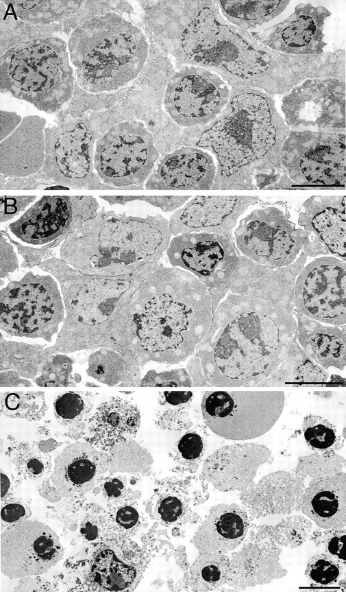
Electron microscopic analysis of livers from E13 Ikkβ+/+, Ikkβ+/−, and Ikkβ−/− embryos. Both E13 Ikkβ+/+ (A) and Ikkβ+/− (B) livers exhibited normal morphology. The Ikkβ−/− liver (C) exhibited varying degrees of apoptosis characterized by collapsed and condensed nuclei and general cellular degeneration. Bars = 5 μm.