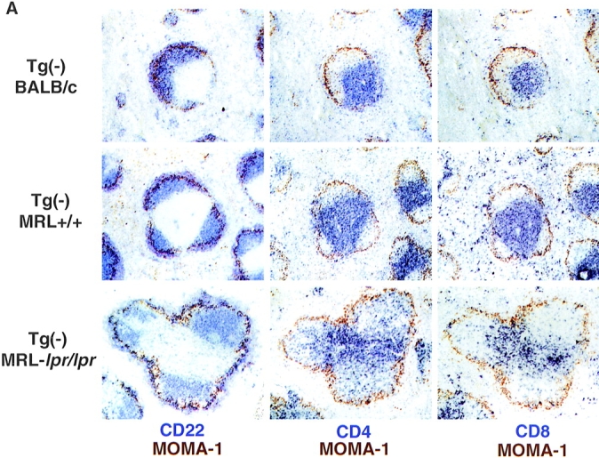
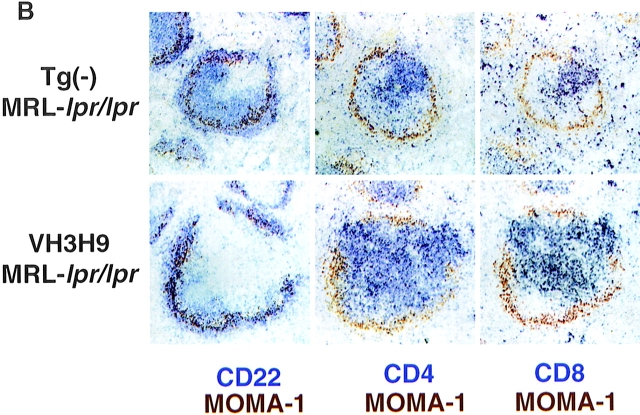
Figure 5.

Disrupted architecture in MRL- lpr/lpr mice. (A) Spleen sections from (top) Tg− BALB/c, (middle) Tg− MRL+/+, and (bottom) Tg− MRL-lpr/lpr mice were stained with Abs against MOMA-1 (MZ metallophilic macrophages) and (left) CD22 (B cells), (middle) CD4 (T cells), or (right) CD8. In MRL-lpr/lpr mice, CD4 T cells are present scattered in the B cell area. CD8 T cells remain in the PALS. Mice shown are 12 wk old. (B) Spleen sections from age-matched (top) Tg− MRL-lpr/lpr and (bottom) VH3H9 MRL-lpr/lpr mice. VH3H9 Tg accelerates the appearance of disrupted architecture in MRL-lpr/lpr mice. Mice shown are 6 wk old. Original magnification: ×100 (n = 15 mice of each genotype).

