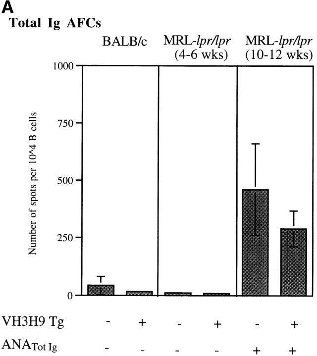
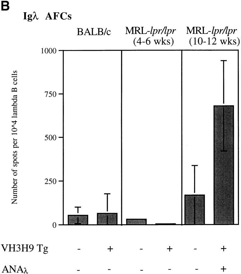
Figure 6.
AFCs in the spleen from ANA− (4–6 wk) and ANA+ (10–12 wk) Tg− MRL-lpr/lpr, VH3H9 MRL-lpr/lpr, and BALB/c mice. The number of (A) total Ig and (B) Igλ AFCs were determined by ex vivo ELISpot. The data are presented as the mean number of AFCs per 104 cells ± the SD of triplicate wells. Total Ig+ and Igλ+ AFC numbers were calculated by normalizing for total B220+Ig+ and B220+Igλ+ numbers as determined by flow cytometry, respectively. Presented data are from a representative mouse of each genotype. Total number of mice from five experiments: n = 5 Tg− BALB/c; n = 4 VH3H9 BALB/c; n = 4 ANA+ Tg− and VH3H9 MRL-lpr/lpr; and n = 2 ANA− Tg− and VH3H9 MRL-lpr/lpr mice. Note that a higher number of Igλ+ AFCs than total Ig+ AFCs were detected for the ANA+ VH3H9 MRL-lpr/lpr mice in this assay. This difference is attributed to a sensitivity difference in the reagents used to detect total Ig versus Igλ. Therefore, although comparisons can be made for the relative number of AFCs of a given type (i.e., between Igλ+ numbers), they cannot be used to compare absolute frequencies between types (i.e., total Ig vs. Igλ).


