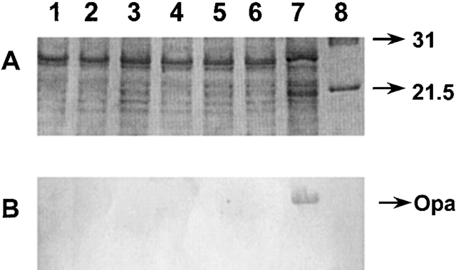
Figure 4.
SDS-PAGE analysis of Opa expression by various N. gonorrhoeae strains. Whole bacterial cell lysates were prepared by suspending bacteria in lysing buffer and boiling the extract for 10 min. An aliquot of the bacterial cell lysate was analyzed on a 13% acrylamide gel using the Tris-glycine buffering system (reference 34). (A) A photoreproduction of the Coomassie blue–stained gel; (B) a Western blot of an identical gel where the proteins were transferred to nitrocellulose and Opa was detected by incubation with the Opa-specific mAb 4B12 (references 42, 43). The lanes represent: 1, F62; 2, F62ΔlgtAlgtG+; 3, F62ΔlgtA; 4, F62ΔlgtD; 5, F62ΔlgtAΔlgtE; 6, F62ΔlgtAlgtC+; 7, PID2; and 8, molecular weight.

