
Figure 8.
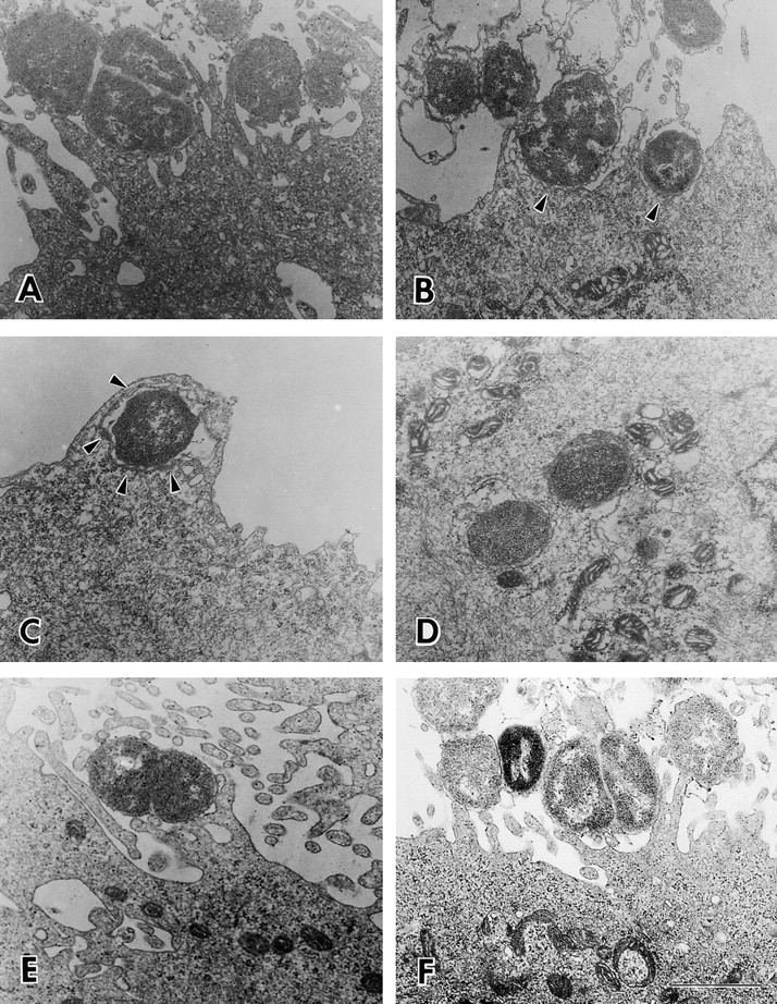
Ultrastructural analysis of ME180 cells infected with gonococci. ME180 cells were inoculated with 107 CFU/ml of gonococci at 37°C for 6 h. The cells were washed, fixed, and processed for electron microscopy. Thin sections (60 nm) were contrasted with uranyl acetate and lead citrate, then analyzed. Shown are representative images. (A–D) The cell inoculated with F62; (E) the cell inoculated with F62ΔlgtA; (F) the cell inoculated with F62ΔlgtAlgtG. Arrows indicate the intimate interaction between gonococci and the plasma membrane of epithelial cells. Bar, 0.5 μm.